


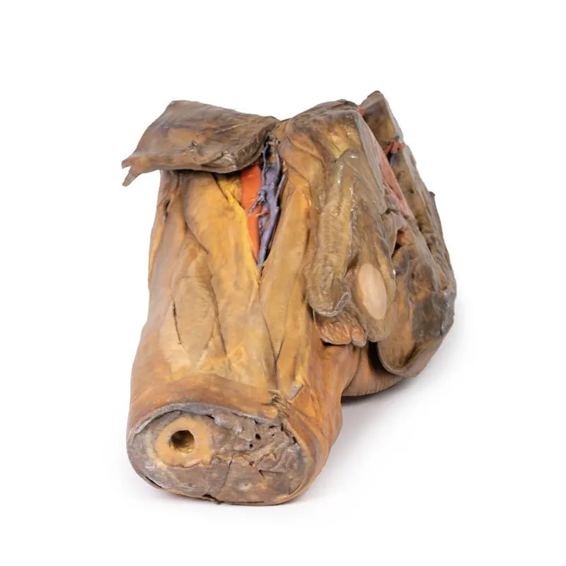
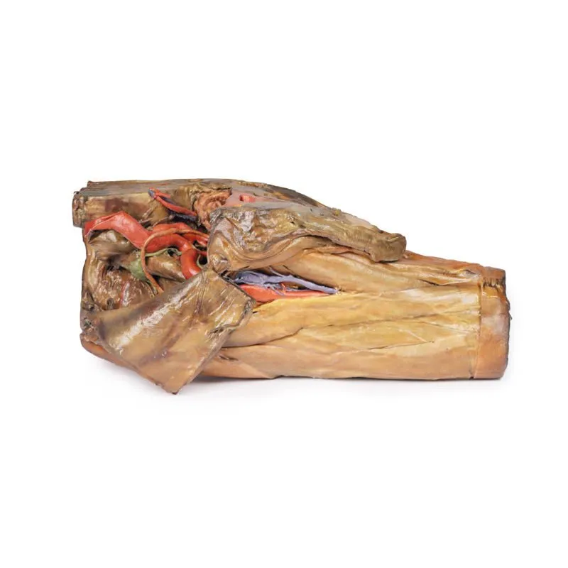

3D Printed Male Hemipelvis and Thigh
This 3D model preserves a right male pelvis sectioned just superior to the L5 vertebra and sectioned at the
midsagittal plane, with the thigh preserved to near the midshaft of the femur. This specimen compliments our LW
91 female hemipelvic specimen and thigh.
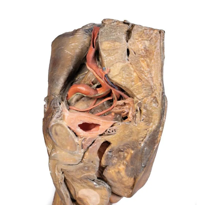
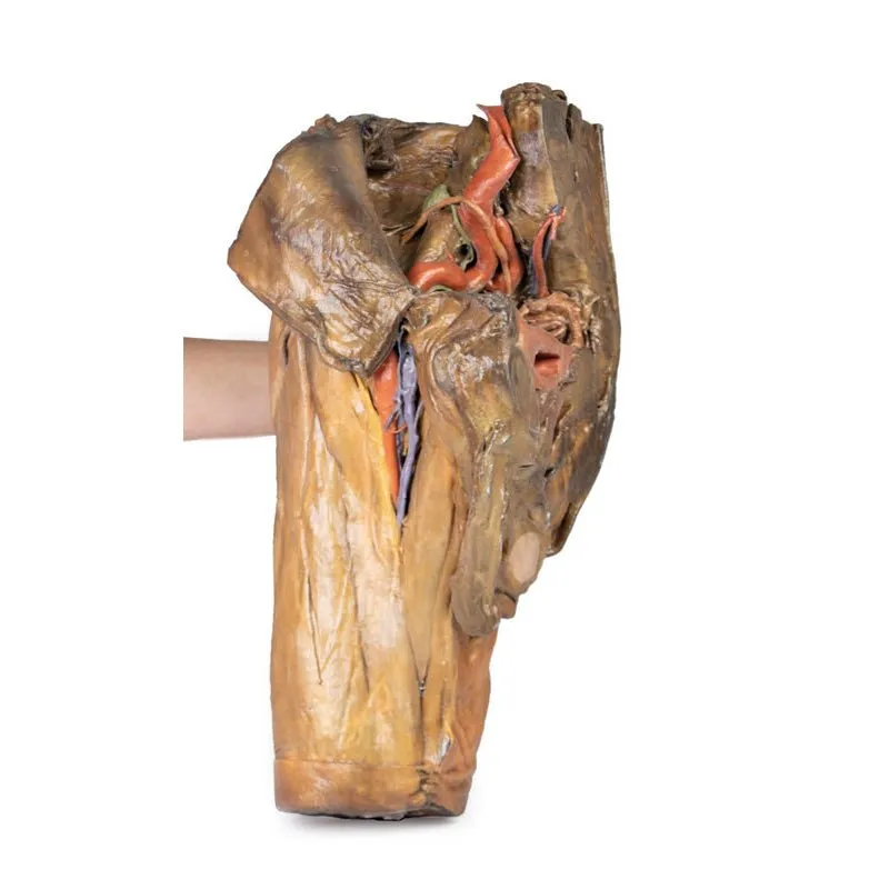
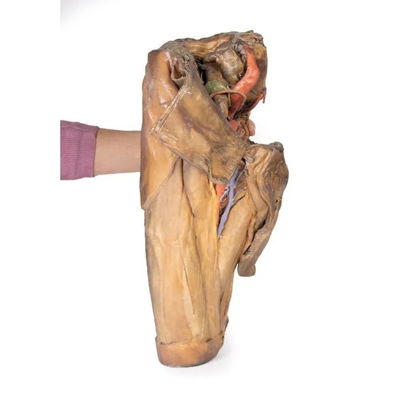
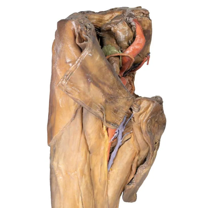
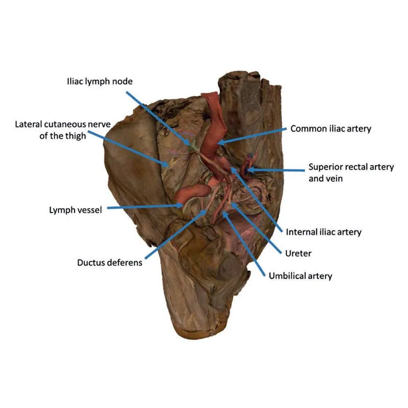
The common iliac artery is preserved with several key branches
visible, particularly the distribution of the internal iliac within the true pelvis. Several major vessels
including the obturator artery and the partially obliterated umbilical artery passes towards the anterior
abdominal wall (to form the medial umbilical ligament) and gives off the superior vesicle artery; while the
roots of the iliolumbar, superior gluteal, inferior gluteal and internal pudendal artery are visible lateral to
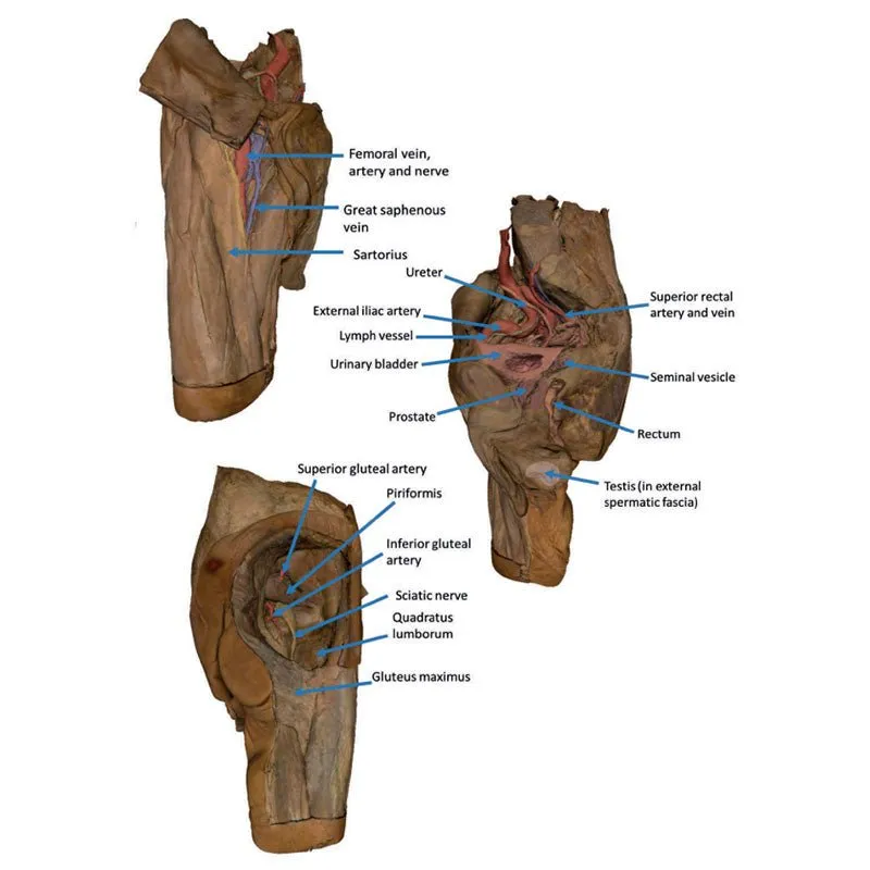
the urinary bladder. The ureter descends superficial to these vessels to approach the urinary bladder which is
covered with peritoneum in this model. The ductus deferens is exposed from the entry into the space via the deep
inguinal ring and passing posteriorly (though sectioned from its normal insertion pathway and resting on the
internal iliac artery). Adjacent to the ureter and on the superficial surface of the psoas major muscle is an
enlarged iliac lymph node and part of the lymphatic vasculature ascending along the external iliac artery. The
majority of the pelvis has been left undissected, allowing for an appreciation of the rectovesicular pouch and
the exposed superior rectal artery and vein approaching the preserved portion of rectum. In cross section, the
rectum, seminal vesicle and prostate are visible (the section plane preserves parts of both the prostatic
urethra and ejaculatory duct).
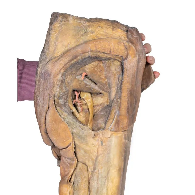
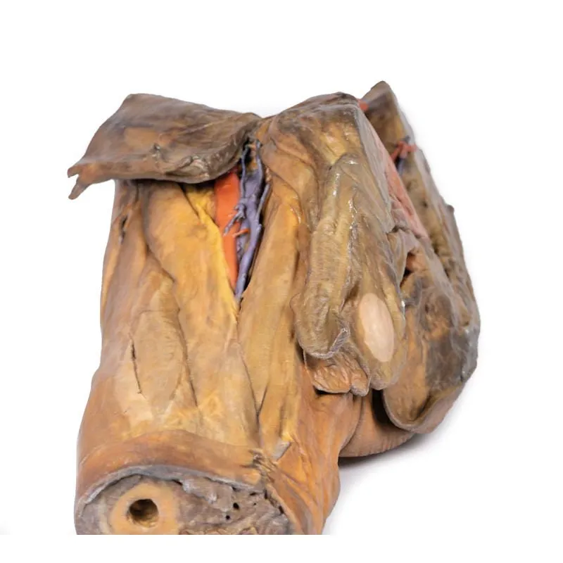
In the anterior thigh the borders and contents of the femoral triangle are well-preserved, with partial coverage by the flap of the anterior abdominal wall. Posteriorly the skin over the gluteal region and the gluteus maximus muscle have been removed as sequential windows to expose the gluteus medius and minimum muscles, the piriformis, the obturator internus with gemelli muscles, and the quadratus femoris muscle. The superior and inferior gluteal arteries are maintained superior and inferior to the piriformis, respectively; with the sciatic nerve exiting inferior to piriformis before passing deep to the retained portion of the gluteus maximus.
Download:GTSimulators by Global Technologies
Erler Zimmer Authorized Dealer


